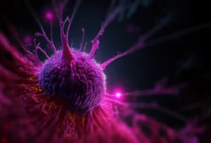
Sample preparation is a critical step in any cell-based workflow, and its success is pivotal in determining the outcomes of downstream processes such as cell growth for patient-derived xenograft (PDX) models, CRISPR transduction efficiency, single-cell RNA sequencing, proteomics, and functional assays like cytokine release assays (Seahorse) or metabolomics studies (Isoplexis)1,2. At the heart of sample preparation lies cell separation, which needs to be thorough, removing all traces of dead or dying cells and debris, and gentle to preserve cell viability, cell representation, and integrity, all while still maximizing laboratory productivity and research ROI. Researchers continue to encounter challenges with existing cell separation methods, and innovation in the sample preparation space has remained stagnant for over three decades.
The current landscape of sample preparation technologies remains dominated by three common methods, however each method presents distinct limitations. As such, it is imperative to consider how innovative approaches to cell separation could address these challenges, thereby improving cell viability, enhancing assay outcomes, and boosting research productivity.
Sample preparation technologies
Researchers across academia and biotech have long relied on traditional sample preparation technologies, which frequently fail to deliver high-quality outputs. This often results in suboptimal assay outcomes or long exhaustive workflows, particularly with challenging samples. The following sections describe the advantages and limitations of these existing technologies.
Centrifugation
Centrifugation is a versatile cell separation technique implemented through gradient centrifugation or wash spins3,4. While wash spins are inexpensive and relatively user-friendly, gradient centrifugation requires time and effort for optimization and is highly dependent on the operator’s skill, which introduces variability in results. Cells are susceptible to damage throughout centrifugation, rendering it unsuitable for particularly sensitive cells like primary neurons. Due to their subjective nature, the decanting, aspiration, and resuspension steps further contribute to the inherent variability and enhance the risk and volume of cell loss. Additionally, this method struggles with handling low numbers of cells, making it less suitable for samples with scarce cellular material, such as precious samples from human subjects5.
Magnetic beads
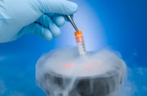
Magnetic bead-based cell separation is another well-established, cost-effective, and widely accessible method known for its ability to specifically enrich different cell types using targeted magnetic beads6. However, it is limited when working with lower cell numbers and larger cell types (greater than 30 micrometers), and column-based methods are often prone to clogging in the presence of excessive debris or dead cells, resulting in impurities in outputs. The process typically involves multiple steps that require significant hands-on time. Moreover, dead cells must be removed separately using annexin-based methods, which can result in further cell loss7. This can be particularly problematic for cryopreserved samples where annexin-based methods can reduce yields significantly.
Flow sorting
Flow cytometry-based sorting is a highly sophisticated tool known for its capacity to sort and analyze cells with high specificity8,9. It requires considerable expertise and is often located in core facilities, requiring users to schedule access and limiting availability for frequent use. Flow sorting uses fluorescent labels to identify specific cell types, which can be toxic to some cell types and restrict cell use in particular assays. Moreover, its use is generally limited to smaller cell types (less than 30 micrometers). Flow sorting relies on gating strategies to isolate populations, which can be complicated by large numbers of dead cells or debris, potentially obscuring rare cell types or causing nonspecific reactivity. Additionally, the high pressure used during sorting may not be suitable for sensitive cell types, posing a risk of damaging the cells.
Rethinking sample preparation methodologies
The limitations of traditional cell separation techniques have significant implications across biomedical research. These limitations require researchers to increase cell numbers to offset the impurities affecting cell counts. Despite these adjustments, cell growth and transduction efficiencies may still be compromised, often resulting in highly variable or inaccurate assay results. Consequently, this can complicate data analysis, render data unclear and unreliable, and may necessitate repeated experiments, thereby consuming valuable time, resources, and funds and causing delays in project timelines. In some cases, these issues can even obstruct the publication of data or hinder the progression of a drug’s development10,11.
Innovation is urgently needed to overcome these issues and their significant implications. The ideal cell separation technology would integrate the following key features:
- User-friendly: Researchers desperately need a simple, intuitive system that can be universally adopted without lengthy, specialized training or the need for access to core facilities, thereby eliminating scheduling delays.
- Flexibility: Given the wide variety of projects conducted within research labs and the ever-changing nature of research, a solution must accommodate a broad range of sample types, including those with low cell numbers or larger cell types, and meet the requirements of specific assays.
- Automated: An optimal cell separation protocol should feature largely automated workflows that minimize hands-on time and reduce variability while enhancing operational efficiency.
- Standardization: Standardization is essential to reproducibility and data validity, and tools should utilize standardized operating procedures to ensure consistent performance across replicates and between users.
- Cost efficiency: Employing cost-effective methods is essential for maximizing return on investment in laboratories with often limited budgets.
A new approach to solving perpetual limitations
LevitasBio® has introduced Levitation Technology™ and the LeviCellⓇ platform as a revolutionary solution to address the limitations of traditional cell separation techniques. By levitating live cells in a paramagnetic solution within a magnetic field, they can be separated from dead or dying cells without labeling, minimizing stress and preserving cell integrity. As a result, the LeviCell® systems enhance the viability and quality of target cell populations, which is particularly beneficial for applications such as CRISPR transductions or PDX models, where it supports researchers in achieving improved efficiencies and better model development.
This improvement in starting material quality enhances the signal-to-noise ratio and ensures that every sample can directly contribute to meaningful data, reducing the need for extensive data filtering or corrections. This is crucial for precision applications like single-cell RNA sequencing, where each cell’s analysis is essential, or drug dosing assays requiring consistent results to accurately evaluate a drug candidate’s efficacy.
The LeviCell system streamlines the cell separation process into three straightforward steps that can be completed in just 20 minutes, significantly shortening timelines, enhancing data validity, and avoiding the waste of resources and research funds. Get in touch today to learn more about how the LeviCell system can support your lab’s research and development needs!
References
1. Capelli C, Cuofano C, Pavoni C, et al. Potency assays and biomarkers for cell-based advanced therapy medicinal products. Front Immunol. 2023;14:1186224. doi:10.3389/fimmu.2023.1186224
2. Niepel M, Hafner M, Mills CE, et al. A Multi-center Study on the Reproducibility of Drug-Response Assays in Mammalian Cell Lines. Cell Syst. 2019;9(1):35-48.e5. doi:10.1016/j.cels.2019.06.005
3. Alberts B, Johnson A, Lewis J, Raff M, Roberts K, Walter P. Fractionation of Cells. In: Molecular Biology of the Cell. 4th Edition. Garland Science; 2002. Accessed April 15, 2024. https://www.ncbi.nlm.nih.gov/books/NBK26936/
4. Harwood R. Cell Separation by Gradient Centrifugation. In: International Review of Cytology. Vol 38. Elsevier; 1974:369-403. doi:10.1016/S0074-7696(08)60930-4
5. Tomlinson MJ, Tomlinson S, Yang XB, Kirkham J. Cell separation: Terminology and practical considerations. J Tissue Eng. 2013;4:204173141247269. doi:10.1177/2041731412472690
6. Plouffe BD, Murthy SK, Lewis LH. Fundamentals and application of magnetic particles in cell isolation and enrichment: a review. Rep Prog Phys. 2015;78(1):016601. doi:10.1088/0034-4885/78/1/016601
7. Hochreiter-Hufford A, Ravichandran KS. Clearing the Dead: Apoptotic Cell Sensing, Recognition, Engulfment, and Digestion. Cold Spring Harb Perspect Biol. 2013;5(1):a008748-a008748. doi:10.1101/cshperspect.a008748
8. Telford WG. Flow cytometry and cell sorting. Front Med. 2023;10:1287884. doi:10.3389/fmed.2023.1287884
9. Ibrahim SF, Van Den Engh G. Flow Cytometry and Cell Sorting. In: Kumar A, Galaev IY, Mattiasson B, eds. Cell Separation. Vol 106. Advances in Biochemical Engineering/Biotechnology. Springer Berlin Heidelberg; 2007:19-39. doi:10.1007/10_2007_073
10. Hajeb P, Zhu L, Bossi R, Vorkamp K. Sample preparation techniques for suspect and non-target screening of emerging contaminants. Chemosphere. 2022;287:132306. doi:10.1016/j.chemosphere.2021.132306
11. Duong VA, Lee H. Bottom-Up Proteomics: Advancements in Sample Preparation. Int J Mol Sci. 2023;24(6):5350. doi:10.3390/ijms24065350