New Tissue Dissociation Workflow for Single Cell Analysis
The complexity of dissociating complex tissue into single cells
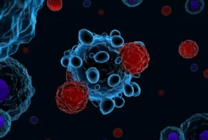
Isolating cells for cancer diagnostics is critical if personalized medicine is to flourish. However, dissociating complex tissues into single cells while maintaining cellular integrity and stable genetic expression is poorly understood and most present approaches in the lab do not have the efficiency that is needed to be clinically relevant for processing of samples of heterogeneous tissue.
A new approach: 15 minute chemical-mechanical dissociation protocol
Writing in a recent issue of Cellular and Molecular Bioengineering, (Vol. 14, No. 3, June 2021 pp. 241–258; https://doi.org/10.1007/s12195-021-00667-y) researchers from the Center for Biomedical Engineering at Brown University reveal a 15 minute chemical-mechanical dissociation protocol for clinically relevant preparation of single-cell suspensions from frozen biopsy cores of complex tissues.
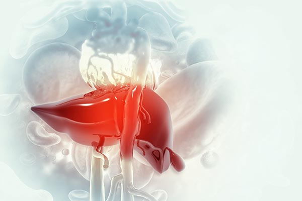
Dissociation and analysis of frozen bovine liver biopsy cores
Frozen bovine liver biopsy cores were normalized by weight, dimension, and calculated cellular composition. Various chemical reagents were tested for their capability to dissociate the tissue via confocal microscopy, hemocytometry and quantitative flow cytometry. Images were processed using ImageJ. Quantitative flow cytometry with gating analysis was also used for the analysis of dissociation. Physical modeling simulations were conducted in COMSOL Multiphysics.
The researchers established that a combination of 1% type-1 collagenase and pronase or hyaluronidase in 100 U/lL HBSS solution is the most effective at dissociating 2.5 mm thawed bovine liver biopsy cores in 15 min, with dissociation efficiency of 37-42% and viability >90% as verified using live MDA-MB-231 cancer cells. Cellular dissociation is significantly improved by adding a controlled mechanical force during the chemical process, to dissociate 93 + 8% of the entire tissue into single cells. The protocol demonstrates that controlled mechanical force in combination with chemical treatment produces high quality tissue dissociation.
Applicability to different tissue and cell types
According to the researchers, the applicability of this workflow is likely to hold for most soft tissues, which are the tissue type that is most frequently dissociated in practice. However, the success of this protocol across different tissue and cell types, especially fibrous, crosslinked, and necrotic or diseased tissues, should be investigated further.

