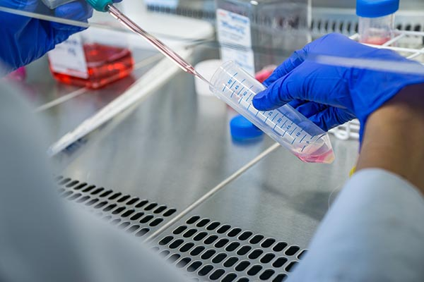
Cells are the basic functional unit of life. Cells of specialized type come together to form a tissue, whereas tissues that perform a specific function compose an organ. Cell culture plays a significant role in studying basic cell biology, cell cycle mechanisms, specialized cell function, cell–cell and cell–matrix interaction. Tissue culture allows for study of cell biology from multicellular organisms in vitro either as 2-D or 3-D cell cultures. Today, advancements in scientific methodology and cell culture matrix reagents provide in vitro environments conducive for 3-D cell culture, where stem cells can be differentiated and grown into organoids, that mimic the cell arrangement of an organ. This blog focuses on the technique of cell culture, 2-D and 3-D, and the importance of tissue dissociation enzymes for cell separation.
Cell Culture vs Tissue Culture: What’s the Difference?
Tissue culture and cell culture are often used interchangeably. So one may wonder, what is the main difference? The source of cells propagated in vitro.
Cell culture is the artificial maintenance of dissociated cells in vitro, either isolated as primary cells, immortalized cell lines or cell strains. This in vitro environment requires supplemental media and growth factors to maintain cell growth. Cell culture studies complement in vivo experiments, as the cells allow for a more controlled environment to assess cellular functions and processes.
Tissue culture is when cells are removed from a parent organism and viable propagation of isolated cells, tissues or organs is maintained in vitro through artificial means. Tissue culture allows for study of cell biology from multicellular organisms. Tissue culture experiments often assess physiological function of cells that make up the tissue and can be used for pharmacological assessment (e.g. cytotoxicology) before moving to in vivo experiments.
What Are Some Common Uses of Cell & Tissue Culture?
Scientists can perform various down-stream assessments from cell culture experiments to decipher mechanisms of action, post-translational modifications and cell-cell relationships. To do these studies, antibodies and cell markers in applications like western blot or flow cytometry can be used to determine protein expression or localization. Furthermore, single-cell applications like immunocytochemistry can also provide the localization of specific proteins in or on a cell, whereas scRNA-seq provides genomic characterization. These types of analyses can help one understand the relationship of regulatory genes and protein expression of a cell. This provides an avenue for understanding cell function when experimental modifications in vitro can be directly correlated with protein or genetic alterations observed at the single-cell level.
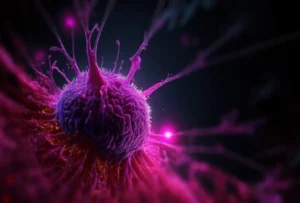
Regenerative medicine using pluripotent stem cells use 3-D cell culture experimental techniques to develop organoids. Organoids derived from stem cells aggregate together to form a spheroid structure that mimics an in vivo organ. To obtain this 3-D structure, extracellular matrices help form the cellular bonds & organization of cells in vitro. Organoids can be used to study human development and disease, drug discovery and perhaps personalized medicine.
What Is Tissue Dissociation?
Tissue dissociation can employ mechanical dissociation, enzymatic dissociation or a mixture of both methods to obtain primary cells from tissue. In short, mechanical dissociation is when a tissue sample is cut, crushed, or pulverized/homogenized using an instrument to get the tissue in smaller, digestible pieces. Mechanical dissociation is often required for tissues that have strong adhesions that need to be broken down before single cells can be extracted. Tissues that undergo harsh mechanical dissociation may result in low viability and low yield of primary cells. A less harsh method of dissociation is mechanical disruption, in which using a pipette (for trituration) or a vortex can help disaggregate the cells from softer tissue with less physical force, improving viability and yield.
Enzyme dissociation, or enzymatic digestion, used in tissue dissociation protocols is important for cell harvesting of primary cells and can often result in the same extraction efficiency as mechanical dissociation. One important factor when assessing your experimental design for enzymatic dissociation is that temperature plays a significant role in enzyme activity. The stability of individual enzymes varies as do their optimal temperature-dependent behavior–where their enzymatic activity is greatest for tissue digestion. Therefore, each experimental protocol requiring tissue dissociation should be optimized based on the tissue type and sensitivity whether it uses mechanical dissociation, enzymatic digestion, or both, to provide optimal primary cell isolation for cell culture.
Specific enzymes used in various tissue digestion processes for cell isolation will be discussed in sections below.
What Are Primary Cells?
Primary cells are directly isolated from fresh tissue of a living organism. Primary cells can be obtained from tissue biopsies or from organ removal. Primary cells are more sensitive than cell lines and require more supplements than classical media. Primary cells are grown and maintained in vitro using various cell culture reagents such as fetal bovine serum, classical medium, supplements, extracellular matrices and antibiotics to minimize contamination growth. Primary cells are not passaged like cell lines and have a finite lifespan in vitro.
What Are The Differences Between Primary Cell Culture and Immortalized Cell Lines (Secondary Cell Culture)?
Primary cells have a limited life span in vitro whereas secondary cells (immortalized cell lines) have an unlimited lifespan in culture with the right conditions. Cell lines are derived from an existing primary cell culture and are created to expand the lifespan and proliferation rate of that cell type. In fact, a main reason cell lines are often utilized in cell culture experiments is that cell lines can be maintained and passaged, providing a consistent experimental cell model.
Secondary cell cultures also tend to be more robust, being less sensitive to in vitro changes than primary cells. Cell lines are also easier to transfect when performing gene engineering experiments, allowing for higher viability after transfection in cell lines than in sensitive primary cells. This experimental flexibility is a benefit for scientists who are investigating specific questions about cellular biology or function at the cellular level for a specific cell type.
However, primary cell cultures are better representations of functional components of tissue compared to immortalized cells as they are a closer match to an in vivo model. Primary cells are more relevant to biomedical research than cell lines. For this reason, there is a growing representation of primary cell culture work performed using applications from basic biology to drug discovery. Tissue dissociation for primary cell isolation is an important technique that is the starting point for primary cell culture. Therefore, understanding how to maintain cell integrity with high viability from inception of your experimental workflow is crucial.
Let’s discuss some limitations of cell culture, what specific enzymes do for cell isolation and how enzyme dissociation of tissue can be optimized for primary cell culture.
What Are Some Limitations of Primary Cell Culture?
Primary cells require more growth time in vitro and have limited growth potential as they will eventually senesce and die. Secondly, the sensitivity of cells can impact cell viability, particularly if one was studying protein expression that is already in low abundance at the single-cell level. These limitations may lead to required repeat experiments or limited time points for analyses. Therefore, it is particularly important to maximize viable cells isolated by enzymatic dissociation of tissue for primary cell culture.
Furthermore, because primary cells need a donor, one may observe an increased variability between experimental groups due to cell cultures created from multiple donor organisms compared to cell lines from a primary cell origin. This may require more experimental vs control cultures (larger n) to allow for observed significant differences. However, a major benefit of using primary cells is that using these cells reduces the expenditure on animal models, making it more cost effective than in vivo work. Primary cells often preface biological findings of in vivo work, thus experimental models are important when choosing to culture primary cells or cell lines, depending on the task at hand.
Enzymatic Dissociation: What Are Some Common Tissue Dissociation Enzymes?
Enzymatic dissociation is the process of using enzymes to digest minced tissue, effectively releasing cells from tissue. Enzyme dissociation is often used to isolate cells from tissue, as it has shown similar efficacy to mechanical dissociation with increased cell viability.
Before adding enzymes, it is important to increase the surface area of the tissue as much as possible through mincing the tissue with instruments like a scalpel, razor blade or scissors. This increased surface area allows the tissue to have increased exposure to the enzyme, while minimizing the amount of time required for digestion. This step minimizes the single-cell’s exposure to the enzyme which further increase rates of viability and maximizes cell dissociation.
Certain enzymes are ineffective alone as a tissue dissociation enzyme (TDE) and often require combinations with stronger enzymes to successfully release cells from the tissue. Specific enzymes are often more effective with certain tissues, so it is best to determine which enzymes are best suited for the tissue you wish to dissociate cells from.
Trypsin, a digestive protease, efficiently induces cell dissociation by breaking down the proteins that facilitate adhesion of monolayered cells to a cell culture dish. However, trypsin shows little selectivity for extracellular proteins and can be ineffective for tissue dissociation when used alone. Trypsin-EDTA solution is a mixture commonly used for tissue dissociation. EDTA is a chelating agent (binds to calcium), not an enzyme, and works to prevent cell aggregation and assists in detaching adherent cells while extending cell viability.
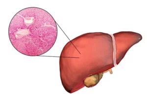
Another common TDE is collagenase, an endopeptidase that breaks down collagen. Collagen is a triple helical protein and the main structural protein in the extracellular matrix and connective tissue that holds tissues together in a body. The triple helix of collagen is resistant to proteolytic cleavage by pepsin, trypsin and papain, whereas collagenase is able to cleave the collagen protein. While there are 28 different types of collagenases, only a handful are most suited for enzyme dissociation of tissues when trying to maximize the number of viable, dissociated cells. Collagenase type 1 is the TDE typically recommended for liver, epithelial, adrenal tissue and adipocytes. Collagenase type 2 can be used for muscle, thyroid, heart, bone, and cartilage degradation.
Elastin, found in elastic fibers of connective tissues, is an extracellular matrix protein. Dissociation of single cells from tissues that have extensive intercellular fiber networks such as lung and kidney tissue require elastase, a serine protease, as the TDE for cell dissociation.
Papain is a cysteine peptidase C1 protease. Papain is a highly efficient TDE that significantly degrades myofibrillar proteins in skeletal muscles and collagen proteins, which hold the tissues together. Papain has been shown to digest neural tissue with greater efficiency and cell viability than other TDEs. Therefore, papain is often used as a neuron isolation enzyme for primary cell culture from central nervous system tissue, such as brain.
DNase is an endonuclease that hydrolyzes phosphodiester bonds and breaks or cleaves either double or single stranded DNA into oligonucleotides. DNase naturally occurs in the cytoplasm of cells, but is often used as a TDE in protocols to digest DNA that leaked out into the buffer as a result of cell damage. There are two classes of DNase; DNase I and DNase II. DNase I can be used on chromatin, double-stranded or single-stranded DNA, while DNase II only cleaves single-stranded DNA. DNase I is often used for reverse transcription experiments, to isolate DNA-free RNA. DNase I is strictly dependent on Ca+2 and is activated by Mg+2 or Mn+2 ions. DNase II is optimally active under acidic conditions and does not require divalent metal ions.
Oftentimes, there are commercially available TDE kits that contain lyophilized enzymes ready to use on your tissue of choice. It is also important to note that enzymes should not be cytotoxic at the concentrations you desire to use them and likely the CoA will notate any testing ranges in cell culture to confirm. When performing tissue dissociation, using the correct TDE is important for maximal harvesting of viable primary cells from tissue.
Whenever working with tissue dissociation enzymes, it is also important to recognize that these enzymes can cleave cell surface markers. This becomes very important for downstream analysis like flow cytometry, western blot or single cell sequencing with cell surface protein analysis. Each of these enzymes cleave epitopes to varying degrees, therefore can affect the detection levels of markers.