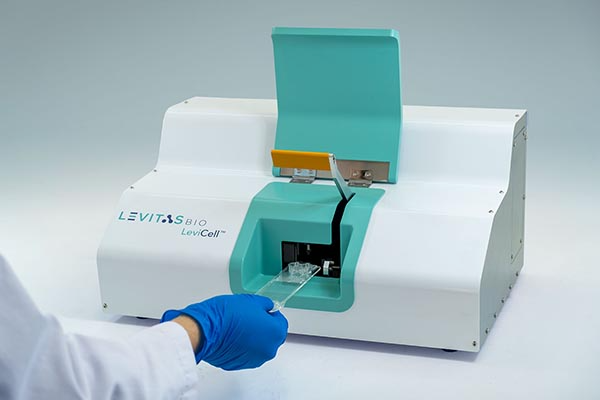
The LeviCell platform from LevitasBio introduces a completely new approach to cell separation that revolutionizes cell enrichment and characterization. Within the LeviCell system, samples such as mammalian cells, bacteria or even viruses are separated using magnetic levitation in a process that is fast, gentle, and minimizes opportunities for perturbation.
We spoke with board member and co-founder Dr. Lars Steinmetz1 about the exciting time at Stanford University when he and colleagues Dr. Gozde Durmus2 and Dr. Utkan Demirci3 initiated a collaboration that revealed the power of magnetic levitation technology when applied to cells, leading to the formation of LevitasBio, and the development and commercialization of the LeviCell.
Dr. Steinmetz also discussed challenges faced by scientists working with cells, how the LeviCell presents a tool to overcome these, as well as opening up entirely new areas of inquiry.
Answers have been edited for clarity.
What is the primary concern or challenge that you see for researchers working with cells, especially for single cell applications, that the technology of the LeviCell might overcome?
There is extensive heterogeneity between cells, and the challenge is to isolate particular cells from complex mixtures. Heterogeneity means that individual cells differ in their properties and their molecular states. For example, researchers may want to isolate healthy cells from a cell culture, or particular cell types from complex tissue samples. Once individual cells are isolated they can be analyzed at a molecular level or used for therapeutic purposes. However isolation of individual single cells is challenging and often not possible with existing technologies.
One needs technologies that separate cells, in a rapid, inexpensive fashion, and with a process that doesn’t perturb the cells along the way. The Levitas system enables us to do inexpensive, gentle, label-free separation of cells in an easy-to-use instrument.
You mentioned one of the advantages of this system is the label-free angle – enabling separation of cells without labels. Why is that so important?
Absolutely, yes. In classical flow cytometry experiments, one often uses surface markers, and antibodies to recognize surface markers with fluorescent labels to demarcate them, and sort them out of a population. But every time you bind a surface receptor on a cell with a molecule, you risk triggering a signaling cascade inside that cell which could ultimately change the cell and its molecular baseline. So, you’re now working with something that has been perturbed and is no longer in its original state.
Therefore, a cell separation technology that doesn’t use any labels is both biologically advantageous and provides a more economical solution – you work with native cells. In the case of the LeviCell, we’re separating based on cellular density, a property that is inherent to any cellular system. Label-free separation also opens up the possibilities dramatically in terms of what you can separate. There aren’t labels available for every cell type or state, so that really limits which cell material you can work on.
However, the LeviCell system doesn’t preclude the usage of labels on top of density-based levitation. Therefore, if a customer wanted to separate two cell populations that have the exact same density, a label can be added to our system to cause a density difference on the cells. The system therefore provides that much needed flexibility.
Is it important that the LeviCell approach to separating based on density is gentler than traditional approaches?
I think that’s absolutely critical. When people separate cells, they often want to do that in a gentle way that preserves cellular state and viability. Cells can often respond to physical stress by changing their gene expression profile so you want to avoid that in any application where live cells are sorted. If you’re just doing cell sorting, because you’re going to extract the genome, then that’s less of an issue. But, in applications where you hope to use the cells, either for therapeutic applications downstream, or you’re going to grow them in the lab, you need the separation to be gentle. Otherwise, again, you’re perturbing the system or potentially even killing the cells.
Could you tell us about the beginnings of LevitasBio?
This whole effort started when Gozde Durmus joined Ron Davis’ and my lab as a post-doc and we started a collaboration with Utkan Demirci’s lab at Stanford. Both Utkan and Gozde are co-founders of LevitasBio. Utkan had developed the instrumentation for magnetic levitation prior to his arrival at Stanford, inspired by seminal work in magnetic levitation from Harvard’s George Whitesides. In Utkan’s group, the technology had previously only been applied to separate biomolecules, and they were just beginning to explore applications in live cells. Utkan and I met at a faculty welcome breakfast and talked about potential opportunities to work together. I mentioned the meeting to Gozde and she was very excited about the idea. Gozde then set out to explore how we could turn magnetic levitation into a sorting technique, and we started collaborating with Utkan’s lab on applying his magnetic levitation technology for the first time to isolate single cells from complex mixtures. When we first saw the potential of the system to enable label-free, gentle, real-time sorting, we really saw an opportunity that we felt was not occupied by any other commercial provider or even any other system that’s in the academic space so far – and that’s how LevitasBio was born.
What types of samples did you first examine using the technology? What types of questions were you asking?
Some of the first things we tried was to test different types of cancer cells, to see whether we could separate them. Gozde took different established cancer cell lines and put them on the Levitas system and we could see the cells separating based on levitation height. When we revisited the equations to calculate levitation height, it turned out that cellular density is the primary driver of the height of levitation above the bottom magnet. This made sense with our data, because we knew that different cancer cells, and even red and white blood cells, have different cellular densities. These initial results then suggested that we could use the system to separate any other cells in complex mixtures as well, as long as they had significant density differences. In the cancer field, we have since explored using the system to collect circulating tumor cells out of blood.
In addition to cancer cells, we have tested bacterial cells in the system, yeast cells in different cell cycle stages and white blood cells that are either activated or not activated. We also tried sickle cells versus healthy red blood cells. Essentially, anything we put onto the system, we were seeing interesting patterns and interesting behaviors. Every time I would meet with Dr. Durmus, she had tried something new on the system, some new cell types, and seen differences. With every new experiment, we discovered something novel, and the potential application opportunities for the system just grew, and grew, and grew.
What do you see as an exciting potential application of the LeviCell?
We’ve talked a lot about the cancer space. I think that’s an exciting area where one would like to isolate rare cells, ideally from the blood of these patients, but these cancer cells are difficult to capture with classical approaches. One typically uses EpCAM surface antibodies to isolate circulating tumor cells from blood. However, as cells go through the epithelial to mesenchymal transition during metastasis, the surface marker EpCAM gets down-regulated, making these cells increasingly more difficult to capture using classical methods. A label-free approach offers inherent advantages here in capturing cancer cells and to then really characterize them in detail.
I think antibiotic susceptibility testing is also another very exciting application. In the lab, Gozde exposed bacteria to antibiotics as they were levitating, and we could show that in real time we could observe changes in their levitation height. When cells were killed by the antibiotic they dropped in their levitation height. So this provides an opportunity to think about doing antibiotic susceptibility testing in a really rapid way. You could imagine that in a doctor’s office, a sample from a patient is collected, processed through the Levitas system to extract the bacterial population, and then in a second levitation experiment, the bacteria are exposed to different antibiotics to observe in real time which antibiotic cocktail actually kills the bacterial population from the patient. These sorts on the Levitas system would only take a few minutes so you could envision making an effective treatment decision while the patient is still in the doctor’s office. This is dramatically faster than the classical antibiotic susceptibility test which requires growing bacteria on antibiotic-containing plates, and then waiting a day or so until colonies either form or don’t form on that plate.
Thinking outside the box
Thinking outside the box in my experience provides the most exciting research breakthroughs. It’s really a fantastic time to play around with magnetic levitation-based density characterization of single cells, and to see what you can do. A lot of opportunities open up when you’ve developed a technology that does something that no other technology has exploited before. In this case it’s the capability of gentle cell separation based on densities without any labels – in a very simple, easy-to-use system. We were just very positively surprised by how many applications we could explore in my lab.
—
Related Publications:
PNAS: Magnetic levitation of single cells
Science: Sorting cells through levitation
References:
1 Dr. Steinmetz is a professor of genetics at Stanford University and Co-director of the Stanford Genome Technology Center, as well as a Senior Scientist at EMBL. In addition to being a member of the scientific founding team at Levitas Bio, he is a co-founder of Sophia Genetics and Recombia Biosciences.
2 Dr. Durmus is an assistant professor of Radiology at Stanford University and a co-founder of Levitas Bio and is currently directing an active research program in exploring various applications of magnetic levitation.
3 Dr. Demirci is a professor of radiology at Stanford University and is currently directing the Canary Center for Early Cancer Detection at Stanford. In addition to being a co-founder of Levitas Bio, he is co-founder of DxNow, Zymot, and Mercury BioSciences.